NEXT STORY
Using 3D electron microscopy to understand viruses
RELATED STORIES
Video URL


NEXT STORY
Using 3D electron microscopy to understand viruses
RELATED STORIES


So then we began looking at electron microscope pictures of various virus particles... plant viruses. Now, we knew what we were looking for but we didn't know... we didn't understand what we were looking at. And I came to realise, what we were looking at was the... was the image projected in... projected in, from three dimensions to two dimensions. Now, people never talked about this. And I remember a man in Oxford, the reader in something or other, a well-known biologist saying to... you know, that they couldn't understand complicated electron micrographs. And I suggested that we actually tilt the specimens... and that came about, tilting the specimen, it was originally, I don't know if you remember, John, I got hold of a... of a... metallurgist stage in electron, which allowed you to tilt the specimens because I thought we should be able to take stereos. And because I can't see with the one eye, I asked people to see what they could see. And nobody could see any stereographic images. And I realised we... there wasn't a stereographic image because that depends upon surface features. And what we were doing was, they were projection, and some way or other the penny dropped and realised that we were looking at a projection. And so the...
[Q] And not just a... a footprint.
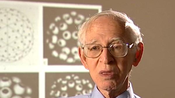
Not just a footprint like X-ray. And so it was quite easy to work out the theory of that. And in fact, taking a series of tilts at different angles is a parallel like collecting X-ray data in a series of two dimensional projections using a camera. I don't think that was explicit in my mind, but really, it was... but before we did this we had been already building models of viruses to explain the pictures purely because we had the theory of how the shells were built and we could explain, for example, as shown there, shown on the slide how you could get very different appearances, very different appearances if you put, say, 180 units into 60 triangles, shown at the top left. And depending on how you cluster the units, we get them clustering in groups of... three, groups of three... that would look like the top right. If you cluster them in groups of six and five they look like that and then you would have on the bottom right clusters of groups of two.
Born in Lithuania, Aaron Klug (1926-2018) was a British chemist and biophysicist. He was awarded the Nobel Prize in Chemistry in 1982 for developments in electron microscopy and his work on complexes of nucleic acids and proteins. He studied crystallography at the University of Cape Town before moving to England, completing his doctorate in 1953 at Trinity College, Cambridge. In 1981, he was awarded the Louisa Gross Horwitz Prize from Columbia University. His long and influential career led to a knighthood in 1988. He was also elected President of the Royal Society, and served there from 1995-2000.
Title: Developing 3D electron microscopy
Listeners: Ken Holmes John Finch
Kenneth Holmes was born in London in 1934 and attended schools in Chiswick. He obtained his BA at St Johns College, Cambridge. He obtained his PhD at Birkbeck College, London working on the structure of tobacco mosaic virus with Rosalind Franklin and Aaron Klug. After a post-doc at Childrens' Hospital, Boston, where he started to work on muscle structure, he joined to the newly opened Laboratory of Molecular Biology in Cambridge where he stayed for six years. He worked with Aaron Klug on virus structure and with Hugh Huxley on muscle. He then moved to Heidelberg to open the Department of Biophysics at the Max Planck Institute for Medical Research where he remained as director until his retirement. During this time he completed the structure of tobacco mosaic virus and solved the structures of a number of protein molecules including the structure of the muscle protein actin and the actin filament. Recently he has worked on the molecular mechanism of muscle contraction. He also initiated the use of synchrotron radiation as a source for X-ray diffraction and founded the EMBL outstation at DESY Hamburg. He was elected to the Royal Society in 1981 and is a member of a number of scientific academies.
John Finch is a retired member of staff of the Medical Research Council Laboratory of Molecular Biology in Cambridge, UK. He began research as a PhD student of Rosalind Franklin's at Birkbeck College, London in 1955 studying the structure of small viruses by x-ray diffraction. He came to Cambridge as part of Aaron Klug's team in 1962 and has continued with the structural study of viruses and other nucleoproteins such as chromatin, using both x-rays and electron microscopy.
Tags: electron microscopy, X-ray, electron micrograph, projection
Duration: 2 minutes, 50 seconds
Date story recorded: July 2005
Date story went live: 24 January 2008
