NEXT STORY
Starting to work on Alzheimer's disease
RELATED STORIES
Video URL


NEXT STORY
Starting to work on Alzheimer's disease
RELATED STORIES


Tony Crowther at this time had started doing electron microscopy from the work on viruses, he actually started doing micro... but I think you, John, taught him how to use the microscope.
[Q] Probably did, yes.
You must have, but I can't remember, did you take pictures of the amyloid?
[Q] No, I think it was Tony.
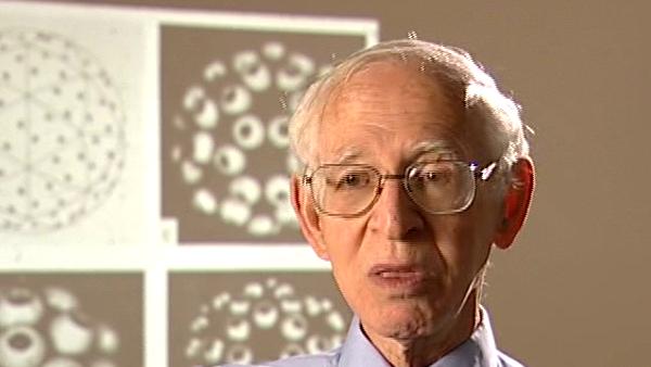
It was Tony Crowther. So Tony Crowther and [Claude] Wischik worked on these fibres and we... and we developed an antibody in Cesar Milstein's group to label these. This was done by a chap called [Michal] Novak working with... so that we could then prove definitely the material that came out of the brains was the paired helical filament. Because you could see it on the microscope labelled by antibodies and at the same time the preparations of course where you could tell the... how much material you extract from the brain by antibody labelling and primulin labelling also was used at the same time. So in the end we were the first people to extract the paired helical filaments, and they did look like filaments in the microscope. Kidd was right, in the sections they did look like a pair, they aren't actually paired helical filaments they're really a single helix, bit like actin, you can describe them in different ways. As a single helix if you look at the... you can think of it in terms of two cross filaments but of course it's a single helix. So at this stage I got quite interested because I did devise the plan of action and I thought, well, now the next thing is to identify the protein, we get it out and the thing to do is to clone... cloning protein had just been introduced. Now there's no cloning going on in structural studies.
Born in Lithuania, Aaron Klug (1926-2018) was a British chemist and biophysicist. He was awarded the Nobel Prize in Chemistry in 1982 for developments in electron microscopy and his work on complexes of nucleic acids and proteins. He studied crystallography at the University of Cape Town before moving to England, completing his doctorate in 1953 at Trinity College, Cambridge. In 1981, he was awarded the Louisa Gross Horwitz Prize from Columbia University. His long and influential career led to a knighthood in 1988. He was also elected President of the Royal Society, and served there from 1995-2000.
Title: Work on Alzheimer's disease: Studying the brain fibres
Listeners: Ken Holmes John Finch
Kenneth Holmes was born in London in 1934 and attended schools in Chiswick. He obtained his BA at St Johns College, Cambridge. He obtained his PhD at Birkbeck College, London working on the structure of tobacco mosaic virus with Rosalind Franklin and Aaron Klug. After a post-doc at Childrens' Hospital, Boston, where he started to work on muscle structure, he joined to the newly opened Laboratory of Molecular Biology in Cambridge where he stayed for six years. He worked with Aaron Klug on virus structure and with Hugh Huxley on muscle. He then moved to Heidelberg to open the Department of Biophysics at the Max Planck Institute for Medical Research where he remained as director until his retirement. During this time he completed the structure of tobacco mosaic virus and solved the structures of a number of protein molecules including the structure of the muscle protein actin and the actin filament. Recently he has worked on the molecular mechanism of muscle contraction. He also initiated the use of synchrotron radiation as a source for X-ray diffraction and founded the EMBL outstation at DESY Hamburg. He was elected to the Royal Society in 1981 and is a member of a number of scientific academies.
John Finch is a retired member of staff of the Medical Research Council Laboratory of Molecular Biology in Cambridge, UK. He began research as a PhD student of Rosalind Franklin's at Birkbeck College, London in 1955 studying the structure of small viruses by x-ray diffraction. He came to Cambridge as part of Aaron Klug's team in 1962 and has continued with the structural study of viruses and other nucleoproteins such as chromatin, using both x-rays and electron microscopy.
Tags: Tony Crowther, Claude Wischik, Cesar Milstein, Michal Novak
Duration: 1 minute, 57 seconds
Date story recorded: July 2005
Date story went live: 24 January 2008
