NEXT STORY

The high-resolution structure of the nucleosome
RELATED STORIES

NEXT STORY

The high-resolution structure of the nucleosome
RELATED STORIES


|
Views | Duration | |
|---|---|---|---|
| 41. Choosing to work on chromatin | 73 | 03:47 | |
| 42. The importance of histones to chromatin structure | 111 | 04:58 | |
| 43. The solenoid model | 123 | 05:44 | |
| 44. Crystallising the nucleosome | 113 | 04:13 | |
| 45. Electron microscope work and crystallising the histone octamer | 81 | 04:16 | |
| 46. The high-resolution structure of the nucleosome | 334 | 06:20 | |
| 47. The solenoid structure and developing new technologies | 1 | 67 | 06:29 |
| 48. Continuing debate on chromatin | 84 | 03:55 | |
| 49. Experiments with lead enzyme | 56 | 04:53 | |
| 50. Work on hammerhead ribozymes with Bill Scott | 175 | 04:52 |


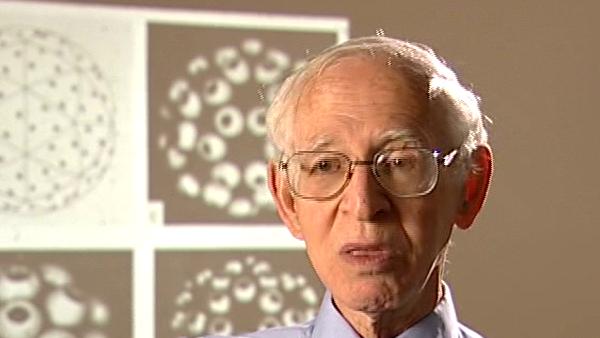
We had done some earlier work at the electron microscope level... which using image processing I'd actually, we had two-dimensional rays. But these... two-dimensional arrays of nucleosomes as well as three-dimensional crystals and we could phase the two-dimensional arrays by electron microscopy and show that they formed a kind of wavy pattern, with a 330 angstrom repeat. And the thing about them is that they looked split into two, they looked bipartite, and this was electron microscopy. And so, eventually you got the crystals and they had a two-fold axis of symmetry parallel to one of the axis, which meant that the nucleosome had... was made up of two equal parts. Now, we knew it was made up of a H3H4 tetramer and 2H2H 2B dimers, so it was fairly obvious what it must be; but we still didn't know the order in which they were. Now, that paper was published in 1977, Finch et al, which was the first one. I don't remember everything that was in that paper, but the two-dimensional work in that paper as well as the three dimensional?
[Q] Think so.
Sorry, I can't remember it all. It was, anyway the work proceeded in stages and in parallel with this... in parallel with this we set out to crystallise the histone octamer and we couldn't get crystals or histone octamer, you needed high salt for this. But they did... the histone octamer was... it formed... it aggregated into helices which you see in an electron microscope. So we did a three-dimensional image reconstruction of the... of the histone octamer and the histone octamer turned out to be a bipartite made of... equal to two-fold symmetry which, as I remember, and Amos used the criteria for checking a two-fold axis and so on. So we... so we therefore had by different methods we had showed that the histone octamer existed and this was without DNA. And it was... it had ridges, in the two fold octamer which you could take 200 base pairs of DNA and wrap them round the ridges.
[Q] Yes.
So therefore we could postulate by different methods and... I spoke about this at a meeting where Hans Zachau was present. And Hans Zachau, he said, 'You haven't got a high resolution crystal structure'; I said, 'I know but we pieced it together from all these different observations.' And then he wanted to know where everything came from, which is a bit of everything because I don't remember whether we had the sequence but there was, in fact, cross linking studies by [Andrei] Mirzabekov, the Russian Group where they found by cross linking studies a very beautiful work... which residues on the H3H4, H2 H2B cross linked to the DNA by cross linking studies. So you could work out the order... along the DNA, the order of... the order of the histones, so I was... we were able in 1980 to postulate a complete structure for the nucleosome with the histones in the right order, all low resolution, all pieced together from different kinds. And that's what drove Hans Zachau a little crazy because he said, 'You haven't got a structure'. I said, 'No, we pieced it together from bits of evidence and this is totally consistent with it'. He said, 'I don't know if he said this, consistency isn't a proof.' And I said, 'You have to produce any other model which explains all these different observations.' So... so that was the histone octamer and the... and the histone... histone octamer and the nucleosome with low resolution.
Born in Lithuania, Aaron Klug (1926-2018) was a British chemist and biophysicist. He was awarded the Nobel Prize in Chemistry in 1982 for developments in electron microscopy and his work on complexes of nucleic acids and proteins. He studied crystallography at the University of Cape Town before moving to England, completing his doctorate in 1953 at Trinity College, Cambridge. In 1981, he was awarded the Louisa Gross Horwitz Prize from Columbia University. His long and influential career led to a knighthood in 1988. He was also elected President of the Royal Society, and served there from 1995-2000.
Title: Electron microscope work and crystallising the histone octamer
Listeners: John Finch Ken Holmes
John Finch is a retired member of staff of the Medical Research Council Laboratory of Molecular Biology in Cambridge, UK. He began research as a PhD student of Rosalind Franklin's at Birkbeck College, London in 1955 studying the structure of small viruses by x-ray diffraction. He came to Cambridge as part of Aaron Klug's team in 1962 and has continued with the structural study of viruses and other nucleoproteins such as chromatin, using both x-rays and electron microscopy.
Kenneth Holmes was born in London in 1934 and attended schools in Chiswick. He obtained his BA at St Johns College, Cambridge. He obtained his PhD at Birkbeck College, London working on the structure of tobacco mosaic virus with Rosalind Franklin and Aaron Klug. After a post-doc at Childrens' Hospital, Boston, where he started to work on muscle structure, he joined to the newly opened Laboratory of Molecular Biology in Cambridge where he stayed for six years. He worked with Aaron Klug on virus structure and with Hugh Huxley on muscle. He then moved to Heidelberg to open the Department of Biophysics at the Max Planck Institute for Medical Research where he remained as director until his retirement. During this time he completed the structure of tobacco mosaic virus and solved the structures of a number of protein molecules including the structure of the muscle protein actin and the actin filament. Recently he has worked on the molecular mechanism of muscle contraction. He also initiated the use of synchrotron radiation as a source for X-ray diffraction and founded the EMBL outstation at DESY Hamburg. He was elected to the Royal Society in 1981 and is a member of a number of scientific academies.
Tags: Hans Zachau, Andrei Mirzabekov
Duration: 4 minutes, 17 seconds
Date story recorded: July 2005
Date story went live: 24 January 2008